

SOX-10
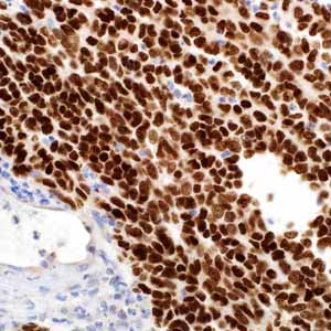
Sry-related HMG-BOX gene 10 (SOX-10) is a nuclear transcription factor that participates in neural crest development and in the specification and differentiation of cells of melanocytic lineage.1 It has been recently shown to be a sensitive marker of melanoma, including conventional, spindled, and desmoplastic subtypes.2 Anti-SOX-10 antibody was applied to a variety of neural crest-derived tumors, mesenchymal and epithelial neoplasms, and normal tissues. SOX-10 nuclear expression was found in 76 of 78 melanomas (97%) and 38 of 77 malignant peripheral nerve sheath tumors (49%), whereas S100 protein was expressed in 71 melanomas (91%) and 23 malignant peripheral nerve sheath tumors (30%).2 SOX-10 was expressed by metastatic melanomas and nodal capsular nevus in sentinel lymph nodes, but not by other lymph node components such as dendritic cells which usually express S100 protein. In scar specimens, immature fibroblasts, epithelioid granulomas, and histiocytic proliferations can histopathologically mimic residual melanoma and even be positive for MiTF and S100.3,4 However, SOX-10 is less likely to be expressed by fibroblasts or histiocytes, especially compared to MiTF and S100. Anti-SOX-10 produces a nuclear stain that provides a clean signal that is much sharper and darker in staining quality when compared to the use of antibodies against MiTF and S100.5 The nuclear signal provided by both anti-SOX-10 and anti-MiTF was easier to interpret than cytoplasmic stains, such as anti-S100 (nuclear and cytoplasmic), anti-HMB-45, and anti-Melan-A (both cytoplasmic), especially in the intraepidermal component where these cytoplasmic markers can non-specifically adhere to melanin. Therefore, anti-SOX-10 has been shown to be superior to all other immunostains in detecting residual invasive and in situ melanoma.1-5 Anti-SOX-10 is also a useful marker in detecting both the in situ and invasive components of desmoplastic melanoma. It is known that the commonly used melanoma markers, anti-HMB-45 and anti-Melan-A, are poorly expressed in desmoplastic melanomas5 while it has been reported that anti-SOX-10 was moderately to strongly expressed in all 28 desmoplastic melanomas tested.2 As an intraepidermal component that is frequently present in greater than 50% of desmoplastic melanomas, the presence of SOX-10 may prove its usefulness over MiTF in the evaluation of melanoma in situ, as it is much more likely to be expressed by a subtle, underlying desmoplastic melanoma than MiTF. SOX-10 is strongly expressed by melanoma cells and is not or very weakly expressed by the spindle fibroblasts in scar.6 These findings underscore the utility of anti-SOX-10 in the differential diagnosis of residual desmoplastic melanoma versus scar and show the clinical value of anti-SOX-10 in the evaluation of melanoma re-excision specimens. SOX-10 is diffusely expressed in schwannomas and neurofibromas. SOX-10 presence was not identified in any other mesenchymal and epithelial tumors except for myoepitheliomas and diffuse astrocytomas.2 SOX-10 expression is seen in sustentacular cells of pheochromocytomas and paragangliomas, and occasionally carcinoid tumors from various organs, but is not seen in the tumor cells.2 In normal tissues, SOX-10 is expressed in Schwann cells, melanocytes, and myoepithelial cells of salivary, bronchial, eccrine, and mammary glands. SOX-10 expression is also observed in mast cells in a variety of tissues and organs in both nuclear and cytoplasmic reaction.