

MUM1 (MRQ-43)
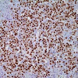
Anti-MUM1 antibody labels a 50kDa, multiple myeloma oncogen-1 (MUM1) protein. MUM1 is encoded by the MUM1/IRF-4 gene, which is mapped to 6q23-25 and identified as a myeloma-associated oncogene1,2. It is a member of the interferon regulatory factor family of transcription factors and plays an important role in the regulation of gene expression in response to signaling by interferon and other cytokines. MUM1 positive cells strongly express the protein in the nucleus in a diffuse and microgranular pattern. However, weak to moderate positivity is also observed in the cytoplasm of MUM1-expressing cells. In normal/reactive lymphoid tissues, such as lymph node, this antibody stains plasma cells, a small fraction of B-cells in the light zone of germinal centers, and a subset of T-cells (5% of T-cells in germinal centers and interfollicular areas)1,2. In non-hematopoietic tissues, anti- MUM1 antibodies recognize normal melanocytes, melanocytic nevi and malignant melanoma1,2. It has been shown that MUM1 protein is expressed in a wide spectrum of hematolymphoid neoplasms and in malignant melanoma. However, it is absent in other human tumors. MUM1 expression has been described in diffuse large B-cell lymphoma (DLBCL) of both pediatric and adult patients. Combined with the immunostaining status of anti-CD10 and anti-bcl6, anti-MUM1 divides DLBCL into 2 main groups: Germinal center type and non-germinal center type (activated B-cell type) with similar 5-year overall survival rates4. Anti-MUM1 antibody can stain other B-cell lymphomas such as lymphoplasmacytic lymphoma, chronic lymphocytic leukemia, follicular lymphoma (mostly restricted in grade 3 follicular lymphoma; negative in low grade follicular lymphoma)3, marginal zone lymphoma, lymphoblastic lymphoma/leukemia, primary effusion lymphoma, primary central nervous system lymphoma, primary mediastinal large B-cell lymphoma, DLBCL, Burkitt-like lymphoma, and classical Hodgkin lymphoma5. However, the tumor cells in nodular lymphocyte predominant Hodgkin lymphoma are negative or only weakly positive in less than 10% of neoplastic cells6. Peripheral T-cell lymphoma (NOS), systemic anaplastic large cell lymphoma (ALK+ or ALK-)7, and CD30+ cutaneous lymphoproliferative disorders8 are the T-cell neoplastic entities that are recognized by anti-MUM1 antibodies4. MUM1 is also strongly expressed in all cases with plasma cell myeloma9. Anti-MUM1 antibody immunostains approximately 92% of malignant melanoma cases, including conventional primary melanoma and metastatic melanoma. However, only a small fraction (14%) of spindle cell and desmoplastic melanomas are immunoreactive with anti-MUM1 antibodies10.