

Adipophilin
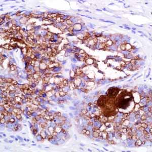
Sebaceous carcinoma is a relatively uncommon cutaneous malignancy and mimics other malignant neoplasms, such as basal and squamous cell carcinomas, and benign processes, such as chalazions and blepharitis, sometimes resulting in delayed diagnosis and suboptimal treatment.1 Adipophilin is present in milk fat globule membranes and on the surface of lipid droplets in various normal cell types. Recently, it has been reported that adipophilin was expressed in 16 of 16 (100%) sebaceous adenomas with a specific pattern: membranous with strong uptake at the periphery of intracytoplasmic lipid vacuoles.2 Of 25 sebaceous carcinomas, 23 (92%) were also labeled with a similar pattern. Additionally, in cases of poorly differentiated sebaceous carcinoma (n=11), in which sebaceous differentiation could not have been reliably interpreted in H&E sections, adipophilin highlighted the sebocytes with a strong membranous labeling of intracytoplasmic lipid droplets in 9 of 11 cases (82%). Moreover, 10 of 10 (100%) xanthelasmas, 9 of 10 (90%) xanthogranulomas, 6 of 6 (100%) xanthomas, and 9 of 13 (63%) metastatic renal cell carcinomas were also weakly-to- moderately positive for adipophilin.2 Expression of adipophilin with a membranous pattern of staining was not seen in any of the other clear cell lesions of the skin, including basal and squamous cell carcinomas, trichilemmomas, clear cell hidradenomas, or balloon cell nevi. Interestingly, a nonspecific granular uptake of anti-adipophilin was seen in adjacent macrophages, keratohyalin granules of epithelial squamous cells, and some tumor cells. Therefore, this anti-adipophilin is suitable for immunostaining formalin-fixed, paraffin-embedded tissue and is helpful in the identification of intracytoplasmic lipids, as seen in sebaceous lesions. It is especially helpful in identifying intracytoplasmic lipid vesicles in poorly differentiated sebaceous carcinomas in challenging cases such as small periocular biopsy specimens.